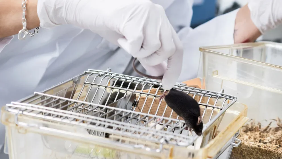
What should you do if your facility is hit with a pinworm outbreak? What impact may this have on your experiments? It’s important that your research mice remain healthy and free from infections, so you can trust your data and reproduce results over time. Luckily, there are many solutions, ranging from treating pinworm-infected mice, to rederivation and re-starting your experiments with “clean” mice.
Do Not Let Pinworms Spoil Your Experiments
Blog Post | February 17, 2015
What should you do if your facility is hit with a pinworm outbreak? What impact may this have on your experiments? It’s important that your research mice remain healthy and free from infections, so you can trust your data and reproduce results over time. Luckily, there are many solutions, ranging from treating pinworm-infected mice, to rederivation and re-starting your experiments with “clean” mice.
What is oxyuriasis?
Oxyuriasis is an infection by one or more species of parasitic nematodes, commonly call pinworms, in the family Oxyuridae. The two species that most commonly afflict mice areSyphacia obvelata and Aspiculuris tetraptera. Pinworm outbreaks occur when the parasites take residence in the gastrointestinal tracts of the animals, and although even low-barrier rooms at JAX are pinworm-free, it is a relatively common occurrence in research facilities worldwide. Pinworm eggs are light, can aerosolize easily, and some are even adhesive-coated. They also become infective within just a few hours after being deposited, and can persist on fomites for extended periods of time. Consequently, they are capable of spreading with ease in both conventional as well as in specific pathogen-free (SPF) facilities, if tight health quality control procedures are not in place.
Pinworm life cycle
The entire pinworm lifecycle, from egg to adult, takes place in the gastrointestinal tract of a single mouse host. Mice become infected directly by ingesting eggs either from the perianal region of an infected cage-mate or indirectly from contaminated materials, such as food, water, bedding, or cage surfaces. Following ingestion, the eggs hatch and larvae establish themselves in either the colon or the cecum, depending on the species, where they mature and replicate.
S. obvelata reside and mate in the cecum or proximal colon while feeding on luminal bacteria. Gravid, adult female pinworms migrate to the perianal area to lay their eggs, which become infectious 5-20 hours after release. The prepatent period (the time between ingestion of infectious larvae to production of eggs) forS. obvelata is 11-15 days, after which the cycle can be repeated in the same host (called retroinfection), or in a naïve, newly-infected animal.
In contrast to Syphacia species, A. tetraptera lay their eggs in the lumen of the colon. The eggs hatch in the cecum, and the larvae migrate to and reside within the proximal colon. After mating, adult females travel to the distal colon to deposit eggs, which are then excreted in feces and become infective 5-8 days later. OnceA. tetraptera eggs become infective, the prepatent period is 21-25 days.
Diagnosis
Pinworm infections don’t typically cause overt clinical signs, although they can cause growth retardation, especially in pre-weanling animals. Diagnosis involves positively identifying pinworm-infected animals by a fecal float test (forA. tetraptera), swabbing, direct examination of colon or cecal contents, or a “tape” test, where cellophane is pressed against the perianal region and immediately placed onto a microscope slide for visualization under a dissecting microscope (forS. obvelata). Because pinworm eggs are intermittently shed in the feces of infected animals, testing feces at multiple time points or from multiple individuals at a single time point is recommended for more effective surveillance. PCR tests also are available, and while they offer increased sensitivity, results may not be as specific as other diagnostic methods.
Treatment
If pinworms break out in your facility, don’t despair! Although it is difficult to eradicate infection, treatments that are coupled with comprehensive room, rack and caging decontamination programs are the most successful. Anti-parasitic drugs, such as fenbendazole and ivermectin, are commercially available, and can be administered topically or in the drinking water (ivermectin) or in feed milled with the drug (fenbendazole). Anthelmintics are not entirely benign compounds, however, and treatment side effects, especially with ivermectin, may occur. Fenbendazole, therefore, may be a safer treatment, not only because it targets all stages of the pinworm life cycle, but because it also has fewer side effects.
If a pinworm infection becomes a persistent problem, or if other pathogens are present in a mouse colony, rederivation is likely the best solution. This process involves in vitro fertilizing oocytes harvested under specific opportunistic pathogen-free (SOPF) conditions to generate SOPF pups. Rederivation can take as little as 12-15 weeks, if done using JAX’s Speed Rederivation service, or between 4-6 months when done via a Custom Rederivation. If you consider the time, cost, effort, and vivarium space necessary to treat pinworm-infected colonies with anti-parasitic drugs, rederivation may be the better option.
Finally, pinworm eggs are hardy, and can persist as environmental contaminants once they are established in a vivarium. Stringent decontamination and frequent swabbing of potentially contaminated surfaces are the best methods to ensure pinworm eggs do not re-infect a colony.
Can it affect my research?
Although pinworm infections are generally considered to be mildly or even non-pathogenic in animals with intact immune systems, they may interfere with research goals in a several ways. Pinworm infections have been shown, for example, to increase a host’s humoral immune responses to nonparasitic antigenic stimuli and to accelerate the development of the hepatic monooxygenase system. In athymic (nude) immunodeficient mice, pinworm infections may instigate lymphoproliferative disorders that may eventually lead to lymphoma. For these reasons, it is important to take appropriate measures to maintain a pinworm-free facility, rederive or treat infected mice, and decontaminate the surrounding environment after an outbreak.
JAX animal health program
JAX maintains strict protocols for the surveillance and monitoring of pathogenic and opportunistic pathogens in all of our facilities. The last pinworm infection at JAX dates back to the 1970’s. Should an outbreak occur, pinworms are one of the pathogens excluded from all JAX mouse rooms, and therefore, all mice shipments would stop to contain the infection.
We realize how important non-infected animals are to investigators’ research, and we are therefore committed to providing the cleanest mice possible from ourhttp://jaxmice.jax.org/health/barrier.htmlMaximum, High, and Standard barriers.
Check out our list of all agents monitored and excluded from JAX facilitieshttp://jaxmice.jax.org/health/agents_list.html
See the JAX Animal Health Reports with pathogen testing results for each animal room
For more detailed information about the procedures used to maintain the high health status of all of our mouse colonies, see our comprehensiveAnimal Health Programhttp://jaxmice.jax.org/genetichealth/health_program.html.
