CD34+ hu-SGM3 Mice

Hu-CD34+ NSG-SGM3 are NSG-SGM3 (NOD.Cg-Prkdcscid Il2rgtm1Wjl Tg(CMV-IL3, CSF2, KITLG)1Eav/MloySzJ, Stock No. 013062) mice engrafted with human cord blood-derived CD34+ hematopoietic stem cells.  NSG-SGM3 mice combine the features of the highly immunodeficient NOD scid gamma (NSG) mouse platform with transgenic expression of human IL3, GM-CSF (CSF2) and SCF (KITLG) cytokines for improved development of human myeloid lineages and regulatory T cell populations.  hu-CD34+ NSG-SGM3 mice exhibit superior, stable engraftment of diverse hematopoietic lineages for immunology and immuno-oncology research.

Body Weights for CD34+ HU-SGM3 Mice

Body Weights for CD34+ HU-SGM3 Mice Body Weights Weeks After Engraftment

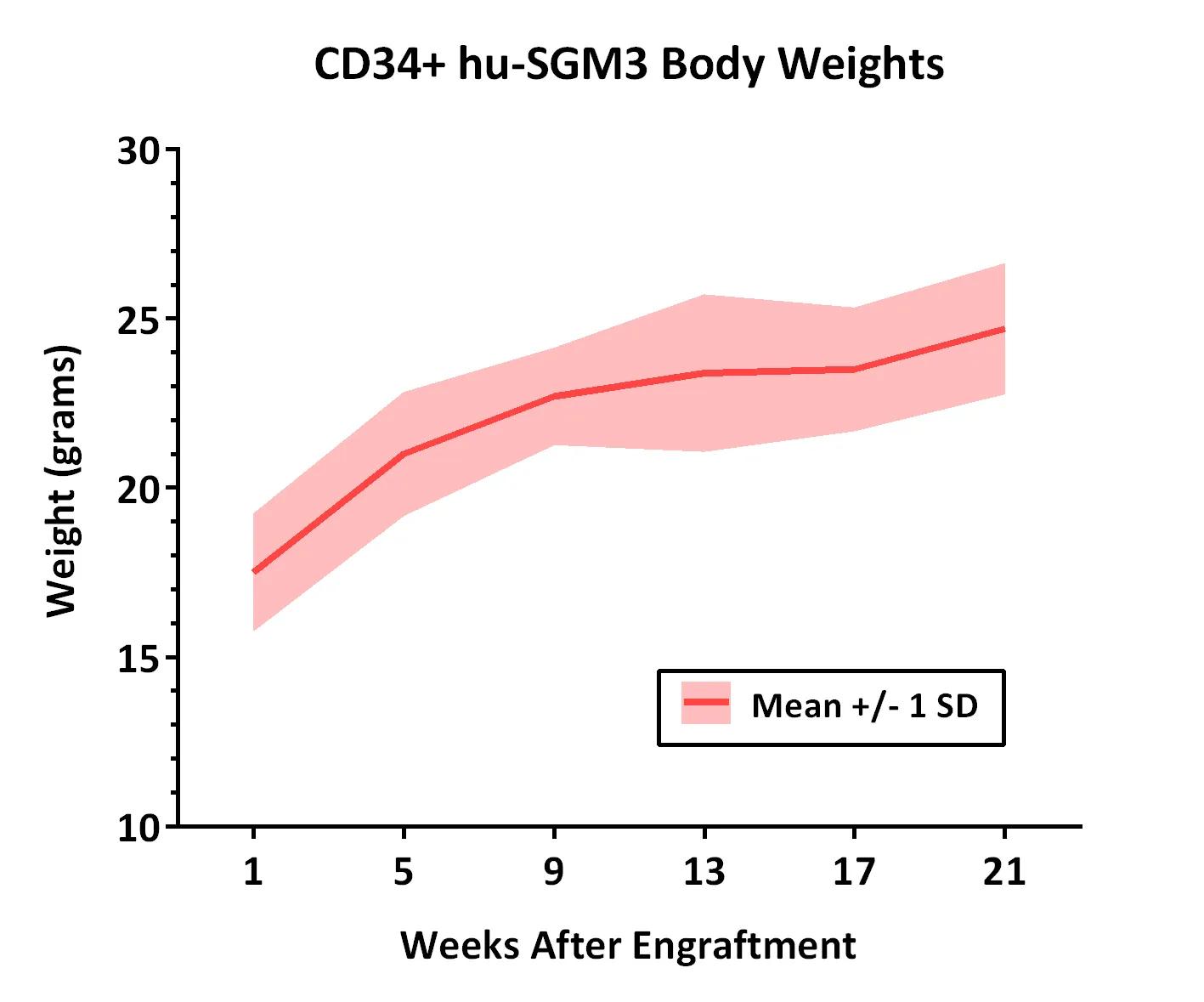
Weights of 10 CD34+ hu-SGM3 mice were measured at different time points after engraftment.  All mice weighed were confirmed to be engrafted with >25% hu-CD45+ peripheral blood cells at the standard time point for analysis (16 weeks after engraftment).  Weights of CD34+ hu-NSGTM mice are also available.

 Weeks After Engraftment 9 13 17 21
 Mean Weight (grams) 17.5 21.0 22.7 23.4 23.5 24.7
 Standard Deviation 1.7  1.8 1.4 2.3 1.8 1.9


Other Considerations

Similar to other immunodeficient strains, maintaining NSG-SGM3 mice in high health status (specific pathogen-free) vivaria promotes overall colony health. The fertility of hu-CD34+ NSG-SGM3 mice has not been tested. The engrafted human immune system is not heritable and would not be transmitted by breeding. A small percentage of hu-CD34+ NSG-SGM3 may develop macrophage activation syndrome (MAS). Please see our FAQ below for more details.

Frequently Asked Questions about Macrophage Activation Syndrome (MAS)

Q) What is macrophage activation syndrome (MAS)?

A) Macrophage activation syndrome (MAS) is a phenotype that occurs occasionally in SGM3 humanized mice generated by transplanting CD34+ human hematopoietic stem cells into the triple transgenic NOD.Cg-Prkdcscid Il2rgtm1Wjl Tg(CMV-IL3,CSF2,KITLG)1Eav/MloySzJ (NSG-SGM3 or NSGS) strain. It is characterized by severe anemia and histopathological evidence of disseminated lymphohistiocytosis, mainly affecting liver, spleen and bone marrow.  MAS is also known as hemophagocytic lymphohistiocytosis (HLH).


Q) What are the clinical signs of MAS in hu-SGM3 mice?

A) Mice with MAS typically present clinically as pale, lethargic, hunched, and in poor body condition. In rare cases, clinical disease may manifest as acute neurological signs, attributed to lymphohystiocytic accumulation within the central nervous system (brain and/or spinal cord).


Q) What factors contribute to the development of MAS?

A) Data collected to date show that MAS occurs in hu-CD34+ NSG-SGM3 due to the expression of human cytokines which promote myeloid cell engraftment and differentiation.
CD34+ donor. The incidence is stem cell donor specific, with approximately 25% of the donors showing increased incidence typically at 15-16 weeks post engraftment and precluding the use of these mice for studies.  In most cases, only a portion of the mice from the same donor will be affected. 
Environment.  It is possible that progression to clinical disease may be exacerbated by exposure to environmental stressors such as suboptimal housing conditions or health status of the facility in which mice are housed. 


Q) Is MAS the same as graft-versus-host disease (GvHD)?

A) This condition is histopathologically, and often clinically, distinct from post-transplantation GvHD, which is characterized by predominant T-cell infiltrates, with damage to the epithelia of the skin, lung, liver, and gastrointestinal tract.


Q) Does MAS develop in other CD34-humanized models like hu-NSGTM?

A) MAS has not been documented in CD34-humanized models involving NSGTM or other related strains. Histiocytosis has been described in some hu-NSGTM mice, although it does not always involve hemophagocytosis and it is not associated with clinical signs of MAS


Q) How can the effects of MAS be minimized in a study?

A) The frequency of MAS varies among CD34+ cell donors for reasons that remain unclear. Consequently, selecting multiple CD34+ donors reduces the risk of loss if one donor becomes affected.  The use of younger mice may be helpful because age is a factor in the development of MAS.


Q) How can a suspected case of MAS be confirmed?

A) If this phenotype is suspected, a veterinarian should be consulted to rule out other conditions specific to immunocompromised mice. Histopathology is the optimal mechanism for confirming MAS, but the facility veterinarian may recommend additional assessments.


Q) Where can I learn more about MAS in hu-SGM3?

A) Relevant publications: Wunderlich, et al., 2016, Yoshihara, et al. 2019, and Janke, et al., 2021.