HLA Expression Improves Immune Response in Humanized Mice
eNews | September 11, 2015Modeling liver infection and human adaptive immunity in mice
Immunodeficient mice engrafted with a human immune system are an important new tool for understanding human-specific pathogens and immune responses to them. Recent research reported in theJournal of Immunology (Billerbeck et al., 2013) demonstrates that expression of human leukocyte antigens (HLA) in the host mouse improves the adaptive immune responses of human immune cells to viral infection of the liver. The authors showed that viral clearance was enhanced due to HLA-mediated, antigen-specific T cell activation.
HLA-expressing immunodeficient mice
Immunodeficient NSGTM mice, NOD.Cg-Prkdcscid Il2rgtm1Wjl/SzJ (005557), can be humanized by engrafting human CD34+ hematopoietic stem cells (hHSCs). Twelve weeks following engraftment, mature human lymphoid and myeloid cells repopulate bone marrow, blood, thymus, spleen and other tissues. One limitation of this model is that immature T cells undergo selection for maturity in the thymus based on antigens presented by mouse leukocyte antigens, and therefore are not capable of human HLA-specific antigen responses. In order to address this issue, Billerbeck et al. created a new NSGTM strain that expresses both HLA class I and II transgenes. NSGTM mice expressing one or the other transgene are available from The Jackson Laboratory; NOD.Cg-Prkdcscid Il2rgtm1Wjl Tg(HLA-DRA*0101,HLA-DRB1*0101)1Dmz/GckRolyJ (012479) and NOD.Cg-Prkdcscid Il2rgtm1Wjl Tg(HLA-A2.1)1Enge/SzJ (009617). The new double transgenic line created by the authors was given the abbreviated name, A2/DR1.
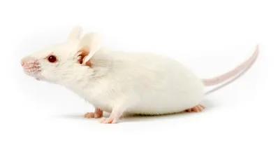
NSGTM mice, stock number 005557 are severely immune-deficient mice capable of humanization, both by breeding in human genes and by transplantation of human cells and tissues.

Billerbeck et al. transplanted NSGTM and A2/DR1 mice with CD34+ hHSCs to establish mice with human immune systems (HIS). The engrafted mice were called HIS and HIS-A2/DR1, respectively. Twelve weeks later, both hosts had similar percentages of CD45+ human leukocytes in blood, spleen, and liver. The two humanized hosts also carried similar percentages of CD3+ T cells, a similar ratio of CD4+ and CD8+ T cells, and similar percentages of NK and myeloid cells. The HIS-A2/DR1 humanized mice, however, showed slightly higher levels of B cells in blood, spleen, and liver compared with the non-transgenic HIS mice. When the mice were treated with PMA/ionomycin, a non-specific immune stimulus, both humanized populations showed increases in human interferon gamma (INFg), tumor necrosis factor alpha (TNFa), and granzyme B, but very little perforin expression. Upregulation of these proinflammatory factors indicates activation of cytotoxic activity by the human T cells. These initial experiments also demonstrated the HIS and HIS-A2/DR1 animals developed very similar human immune cell populations following engraftment of CD34+ hHSCs.
A2/DR1 hosts have an improved functional immune response to hepatic viral infection
The next question the authors addressed was whether the humanized HIS-A2/DR1 mice were capable of mounting improved immune responses compared to the non-transgenic, humanized HIS mice. They were specifically interested in their responses to hepatotropic infections. Adeno-associated viruses (AdV) efficiently infect mouse liver and initiate strong immune responses in mice that have a normal immune system. Billerbeck et al. infected mice with an AdV5 (serotype 5) virus engineered to be replication incompetent. The virus was also modified to express luciferase, which allowed the kinetics of infection to be followed in live mice by bioluminescent imaging. Normal, immune-competent BALB/cJ (000651) mice infected with the virus cleared it from their livers within 10 days. NSGTM mice transplanted with allogeneic bone marrow from C57BL/6J (000664) mice also cleared the virus by 20 days. Strikingly, neither HIS nor HIS-A2/DR1 mice were able to clear the AdV5 infection by 20 days. HIS-A2/DR1 mice, however, did show decreased liver photon flux compared to the HIS mice at both 10 and 20 days, indicating improved immune function due to HLA expression. This initial conclusion was supported by a second infection experiment, in which the mice were pretreated with antibodies to deplete T cells. Indeed, the T cell-depleted HIS-A2/DR1 mice, but not the non-transgenic HIS mice, had a significantly diminished ability to clear the viral infection. Also, the livers of non-T cell depleted HIS-A2/DR1 mice had increased T cell infiltration compared to HIS mice, and a high percentage of these T cells were CD8+ and CD45RO+, which is identified them as effector cells. Further characterization of these T cells from the livers of HIS-A2/DR1 mice revealed that between 10 and 20 days post infection, they declined in CD127 expression and increased in PD1 expression. CD127 is expressed primarily on naïve T cells and is down-regulated on effector T cells. PD1 expression increases during chronic infection. Taken together, this data indicated that antigen presentation by HLA enhanced the immune reactivity towards the viral infection and that human T cells mediated the response through antigen recognition.
The combination of humanization by HLA expression and transplantation of hHSC in the NSGTM mice establish a new and robust platform to better understand the immuno-biology of hepatotrophic infections. The platform will hopefully allow development of new treatments for these very serious diseases.
Additional Research Resources Available:
- Review breakthrough research using NSG mice
- Learn more about humanized NSG mice and request some today
- Discover how In Vivo Pharmacology Services can accelerate your research
Reference
Billerbeck, et al. 2013. Characterization of human antiviral adaptive immune responses during hepatotropic virus infection in HLA-transgenic human immune system mice.J Immonol. Aug 15;191(4):1753-64. PMID: 23833235
