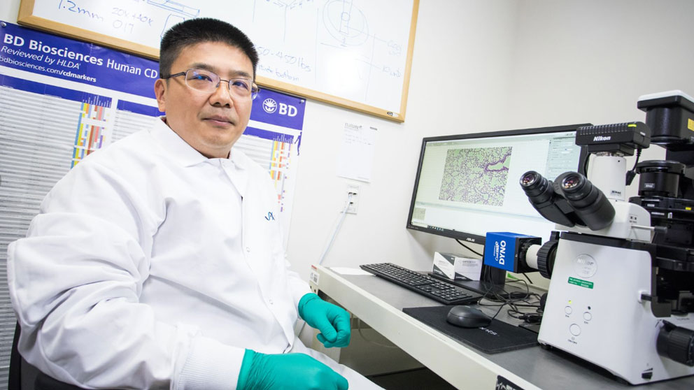
 Photo credit: Tiffany Laufer
Though damaging in itself, the primary tumor that forms after the initiation of cancer is not usually lethal. More dangerous are the cells that separate from the tumor, travel through the bloodstream, and take root elsewhere in the body, in a process known as metastasis that accounts for the majority of deaths from solid cancers.
Fortunately, most of these cells are eliminated by the body’s own immune surveillance mechanism, which kills abnormal and mis-located cells. But how do the cells that establish metastatic cancers escape and survive in a distant, hostile environment?
Jackson Laboratory (JAX) Assistant Professor Guangwen “Gary” Ren, Ph.D., studies metastatic cancers and their interactions with the immune cells populating their new environments. Much attention has been focused on the pro-tumor functions of cells in the innate immune system, known as myeloid cells (macrophages, neutrophils, monocytes) that usually provide swift, efficient clearance of pathogens and malignant cells. But why, in metastatic cancer, do they fail to do their jobs? In “Lung fibroblasts facilitate pre-metastatic niche formation by remodeling the local immune microenvironment,” published in Immunity, Ren and colleagues, including JAX Professor Lenny Shultz, Ph.D. and Assistant Professor Lucas Chang, Ph.D., show that the structural cells in the lung—fibroblasts—drive myeloid cell dysfunction to create a more welcoming environment for metastatic cancer.
Metastatic cancer microenvironment in the lung
Among other functions, fibroblasts are important maintenance cells, contributing to the formation of connective tissues, maintaining tissue structure and helping with wound healing. The latter process involves complex interactions between fibroblasts and immune cells, and while their possible importance has been extensively studied in immune diseases, their roles are not well understood in cancer. The lung is a common site for metastasis, so Ren and his team investigated why it might provide a relatively permissive growth environment. They identified a population of lung fibroblasts that express high levels of genes associated with inflammation. Not only do these particular cells interfere with the immune function of various myeloid cells, the researchers found that they can reprogram them to actually suppress adaptive immune response.
Even before the initiation of metastasis, the fibroblasts affect the function of dendritic cells, the immune cells that trigger anti-tumor adaptive immunity by stimulating T cell production and activation. The research team evaluated a variety of cell types in the lung and found that fibroblasts producing high levels of an enzyme called cyclooxygenase 2 (COX-2) inhibited the expression of dendritic cell genes associated with their ability to identify and present specific targets (antigens) to T cells for tumor elimination. In addition, the expression of genes associated with immunosuppression was found to increase in both dendritic cells and monocytes upon their contact with lung fibroblasts.
COX-2 is involved in the production of the prostaglandin PGE2, a type of physiologically active lipid. It is PGE2 that is secreted by the fibroblasts, affecting immune function in the lung microenvironment around them even in steady state conditions. Interestingly, when the researchers evaluated fibroblasts from other tissues, they found that the situation is unique to the lung: lung fibroblasts produce a higher level of PGE2 than fibroblasts in other tissues. Furthermore, in tumor-bearing mouse models, they found that the regulatory effect of the fibroblasts on myeloid cells was strengthened through IL-1b, a pro-inflammatory signaling molecule mainly produced by myeloid cells. Thus, progression of tumor-associated inflammation makes the lung microenvironment even more receptive to metastatic cancer cells.
Making the surroundings more hostile to cancer
COX-2 is produced from a gene called Ptgs2, so to see what happens when it is absent, Ren and his team developed a mouse model to eliminate Ptgs2 specifically in fibroblasts. As expected, steady-state lung immune cell function and activity increased in these mice. When metastatic breast tumor cells were transplanted into both wild-type (normal) and Ptgs2-lacking mice, the growth of the primary tumor remained unaffected, but metastatic spread of the cancer cells to the lungs was significantly lower in the Ptgs2-lacking mice. Furthermore, combining a COX2-PEG2 blockade with anti-PD-1, a common cancer immunotherapy, was far more effective than either regimen alone. Indeed, the combination reduced the metastatic burden in the mice about 20 times compared with untreated control mice.
Metastatic cancer is still a diagnosis that often comes with a poor prognosis for the patient. Understanding how metastasis occurs and why the lung is such a permissive environment for metastatic cells is an important step toward developing better therapeutics. Because lung fibroblasts actively reconstruct the immune landscape in the lung, targeting fibroblast-associated signaling is a promising new way to treat lung metastasis, particularly in combination with immunotherapies that are already in wide use.