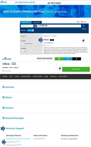
Genotyping Resources

Genotyping protocols based on visible phenotype

B6EiC3Sn a/A-Gy/J mice (stock number: 000503)

Genotyping protocol for Gy mice (August, 2001)

The Jackson Laboratory utilizes visible phenotype to genotype animals from the B6EiC3Sn-a/A-Gy (Stock Number 000503) colony. Animals carrying the allele display a circling behavior whereas animals do not.

The following reference provides PCR assay information that may be used to genotype animals. We do not have experience using the protocols, but offer them as potential alternatives to phenotyping methods.

Strom TM, Francis F, Lorenz B, Boddrich A, Econs MJ, Lehrach H, Meitinger T. 1997. Pex gene deletions in Gy and Hyp mice provide mouse models for X-linked hypophosphatemia. Hum Mol Genet 6:165-171.

B6.Cg-PhexHyp/J (stock number: 000528)

The Jackson Laboratory used to use visible phenotype to genotype animals from the B6.Cg-PhexHyp/J (stock number 000528) colony. Animals carrying the PhexHyp allele have reduced body size with shortened hind limbs and tail whereas animals with the Phex wild type allele do not.

Lepob mice (stock numbers: 000632 and 000696)

The Jackson Laboratory utilizes a combination of breeding scheme and visible phenotype to genotype animals from its B6.Cg-Lepob (stock number 000632) and BKS.Cg-Lepob/+ (stock number 000696) colonies.

A cross between B6129PF1-Aw-J/Aw females transplanted with Lepob/Lepob homozygous ovaries and C57BL/6J males produces heterozygous offspring. Homozygous (Lepob/Lepob) progeny from matings of these Lepob/Lep+ heterozygotes are identified phenotypically by their obesity as early as ~4 weeks of age.

Lep ob genotyping protocols:

  • Lep ob, end point analysis 
  • Lepob, pyrosequencing 
  • Lepob, restriction enzyme digest

Genotyping protocol based on protein staining

Lac-Z (β-galactosidase) activity staining

Lac-Z detection in tail biopsies

  1. 2mm tail biopsies are collected in 1X PBS (see recipes below) in a 1.5ml microfuge tube. Be sure to include a known +/+ (lac-Z negative) biopsy as a control.
  2. Replace 1X PBS solution over biopsy with 2.5% Glutaraldehyde diluted in SpM buffer (see recipes below).
  3. Pre-warm microwave by placing 300ml H2O in a beaker in microwave and heating on "high-power" setting for 30 sec. Remove from microwave.
  4. Microwave biopsy 7-10 sec. with microfuge tube lids open.
  5. Wash biopsy 3 times in 1X PBS.
  6. Replace final PBS wash with 200-500ul staining medium (see recipes below); enough to cover tissue completely.
  7. Incubate @ 37°C 1-3 hrs (alternatively the incubation can proceed overnight at room temperature).
  8. By 15 min. of incubation blue color should be visible at the exposed biopsy surface (where the tail was cut).
  9. Stop reaction by replacing staining solution with 1X PBS. These may be stored at 4°C for several weeks. During this time color deposition will begin to leach into solution.

Reagents:

1 X Phosphate buffered saline (PBS)

0.2g                 KCl

0.2g                 KH2 PO4

1.15g               Na2HPO4

8.0g                NaCl

to 1 liter with DDI H2O

1X SpM

2mM                  CaCl2

2mM                  MgCl2

0.3%                 Glucose

0.3%                Fructose

made in 1X phosphate buffered saline

Staining medium (WEAR GLOVES and WORK IN A HOOD)

5mM                  potassium ferricyanide

5mM                  potassium ferrocyanide

1 mg/ml            X-gal (in dimethyl sulfoxide) final concentration

0.1M                Hepes (pH 7.4)                                                  

made in 1X SpM

Adapted from:

Murti, JR and Schimenti, JC. "Microwave-Accelerated Fixation and Lac-Z Activity Staining of Testicular Cells in Transgenic Mice". Analytical Biochemistry :198, pp. 92-96 (1991).

Genotyping protocols for other strains

BALB/cJ-Trfhpx/JUthHmsJ mice (stock number: 004546)

Trfhpx sequence:

AGGAGCGGAGTACATGCAATCTGTCGGTAACATGAGGAAGTGCTCAACCTCAC(g/a)TAAGTAGAATGAACCCTGTGGAGAGGTCGCAGAGAAACCCAGCGATGGGTGCTGGAATGGTAGTTACAAGAACTCAAACCCTTAGGAAACAGC

Mutant = A
Wild type = G

Genotyping protocol provided by the donating investigator of strain BALB/cJ-Trfhpx/J  has provided this genotyping protocol as a courtesy. The Jackson Laboratory has not used this protocol.

Primers

F 5' tcaagtccaccaccaaggacc 3'

R 5' ctgggtttctctgcgacctctc 3'

The amounts of primer solution correspond to ~20 pmol of each primer. Genomic DNA was prepared using the Qiagen kit and eluted in 200 ul of water.

Cycling conditions

  1. 94oC 3 min
  2. 94oC 40 sec
  3. 61oC 45 sec
  4. 72oC 40 sec
  5. repeat steps 2-4 39 times
  6. 72oC 7 min
  7. 4oC

Clean amplified fragment using phenol/chloroform/iso-amyl alcohol mix (25:24:1). Resuspend in 10 ul of miliQ water.

Restriction digest

The enzyme to be used is Hyp CH4 IV (NEB), an isoschizomer of Mae II. This enzyme works in low salt concentrations and acts poorly on samples taken out straight from the PCR tube. Run samples in a 1.5% agarose gel.

Analysis

  1. Hyp CH4 IV cuts DNA amplified from a wild-type animal. There are two fragments sizes: ~139 bp and ~45 bp.
  2. Hyp CH4 IV does not cut DNA amplified from a homozygous animal (the restriction site has been destroyed by the mutation). There is one full-length band ~184 bp.
  3. The heterozygous animal has all three bands.

PCR resources

Typical 25 µl PCR reaction:

1-5 µl DNA 2.5 µl 10x Perkin Elmer buffer, 1.5 mM MgCl2 (final) 2 µl 2.5 mM dNTP mixture (2.5 mM each dNTP, 200 µM final) 0.5 µl 20 µM forward primer (0.4 µM final) 0.5 µl 20 µM reverse primer (0.4 µM final) 0.1 µl Taq DNA polymerase (can decrease to 0.05 µl) dH2O to 25 µl

(Adapted from Higuchi, R. (1989) Amplifications 2: 1-3)

Quantitative (real-time) PCR (qPCR)

Method that quantifies DNA sequences by normalizing to a reference gene, or to the amount of DNA in the reaction. QPCR is the easiest way to differentiate homozygous (Tg/Tg) from hemizygous (Tg/0) transgenic mice or to determine transgene copy number.

For more information:

Melting curve analysis

Method that uses the melting point of double-stranded PCR products to determine the size of the DNA fragments amplified for transgenes and knock-ins (eliminating the need for agarose gels). This requires a fluorescent dye (typically SYBR green) and special thermocycler (e.g. LightCycler® 480 from Roche). Melting curve analysis protocols provided on JAX® Mice datasheets can be used as a starting point for standard PCR, by eliminating the SYBR green and optimizing the protocol for use in a standard thermocycler.

Pyrosequencing

Sequencing method that uses a chemiluminescent enzyme to detect specific nucleotide incorporation into DNA to genotype knockout and spontaneous mutants. For more information:

  • Pyrosequencing (You Tube)
  • Fakhrai-Rad et al. (2002). "Pyrosequencing: an accurate detection platform for single nucleotide polymorphisms". Hum Mutat. 19: 479.  PMID 11968080

References for PCR

  • Erlich HA.  1989.  Polymerase chain reaction.  J Clin Immunol.  9(6):437-47. Review.  PMID: 2698397.
  • Erlich HA, Gelfand D, Sninsky JJ.  1991.  Recent advances in the polymerase chain reaction.  Science.  252(5013):1643-51.  PMID: 2047872.
  • Henegariu O, Heerema NA, Dlouhy SR, Vance GH, Vogt PH.  1997.  Multiplex PCR: critical parameters and step-by-step protocol.  Biotechniques. 1997 Sep;23(3):504-11.  PMID: 9298224.
  • Truett GE, Heeger P, Mynatt RL, Truett AA, Walker JA, Warman ML.  2000.  Preparation of PCR-quality mouse genomic DNA with hot sodium hydroxide and tris (HotSHOT).  Biotechniques.  29(1):52, 54.  PMID: 10907076

Books on PCR

  • "PCR Technology:  Principles and Applications for DNA Amplification." Erlich HA  (ed.) New York:  Stockton Press, 1989.
  • "PCR Protocols: A Guide to Methods and Applications." Innis MA, Gelfand DH, Sninsky JJ and White TJ (eds.) San Diego, CA:  Academic Press, 1989.
  • "PCR" McPherson MJ, and SG Moller.  New York: Springer-Verlag New York, Inc., 2000.