Spinal muscular atrophy (SMA) is an autosomal recessive disease and the leading genetic cause of infant and toddler death worldwide. It is characterized by the loss of motor neurons in the spinal cord, leading to the inability to control voluntary muscle movements. Affected children are weak, cry feebly, and have trouble swallowing, sucking and breathing. There is no cure, but two recent studies have shown that either restoring or increasing the production of the spinal motor neuron (SMN) protein markedly alleviates or even reverses SMA in mice, offering hope to SMA-affected children and their parents.
SMA
SMA is caused by the absence of the survival motor neuron 1 (SMN1) gene on chromosome 5. The gene's encoded protein, SMN, is evolutionarily conserved, ubiquitously expressed, and essential for localizing and processing subcellular RNA. Due to an evolutionarily recent duplication, humans have two nearly identical copies of the SMN gene – SMN1 and SMN2. Although people with SMA have no SMN1 gene, they have at least one copy (sometimes several copies) of the SMN2 gene. Generally, the more SMN2 copies they have, the less severe is their SMA. Based on disease severity and age of onset, SMA is subdivided into three types: type 1 (severe), type 2 (intermediate) and type 3 (mild). The absence of both the SMN1 and SMN2 genes is embryonically lethal. Although there is no effective therapy for SMA, scientists have speculated that increasing SMN protein production from one or more SMN2 gene copies might compensate for the absence of the SMN1 gene and mitigate, prevent or even cure SMA. Indeed, Smn1-deficient mice that are transgenic for eight copies of the uniquely human SMN2 gene do not develop SMA.
‘Milk’ hormone alleviates SMA in mice

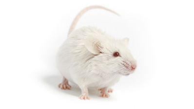
Several molecules had been known to induce SMN2 production in mouse and human cell cultures, putatively via STAT5 signaling, but these molecules can't pass the blood-brain barrier. In contrast, the hormone prolactin, a known STAT5 inducer, can pass the barrier, and its receptors are present in motor neurons. Researchers led by Dr. Alex MacKenzie from the University of Ottawa, Canada, and Faraz Farooq from the Universidad de Granada, Granada, Spain, hypothesized that prolactin could be used to induce SMN2 production and mitigate SMA severity. The MacKenzie/Farooq team tested their hypothesis in a series of experiments in cell cultures and in SMA mouse model FVB.Cg-Tg(SMN2*delta7)4299Ahmb Tg(SMN2)89Ahmb Smn1tm1Msd/J (SMAΔ7, 005025) (Farooq et al. 2011).
By administering prolactin to NT2 and/or MN-1 cell cultures, the MacKenzie/Farooq team demonstrated that prolactin increases SMN production, verified that it both increases STAT5 levels and activates the STAT5 pathway, and showed that it stimulates SMN production via STAT5 signaling.
In vivo experiments substantiated prolactin's role in increasing SMN levels. The MacKenzie/Farooq team demonstrated that prolactin dose-dependently stimulates SMN production in the brain and spinal cord of CD-1 mice, significantly increases SMN2-derived full-length SMN transcripts and protein levels in the brain and spinal cord of SMAΔ7 mice, significantly increases SMN protein levels in motor neurons of the brain and spinal cord and in endothelial cells of SMAΔ7 mice, mitigates weight loss, improves motor function, significantly extends the lifespan of SMAΔ7 mice (21 vs 14 days in non-treated controls), and results in significantly higher SMN protein levels at the time of death in the brain, spinal cord, and muscle of SMAΔ7 mice compared to controls.
In summary, the MacKenzie/Farooq team demonstrated that prolactin increases SMN protein production via STAT5 signaling, has significant therapeutic effects in an SMA mouse model, and is a potentially promising treatment for human SMA.
Restoring SMN production reverses SMA in mice
Although several researchers had examined strategies for treating SMA by increasing SMN production, none had examined the disease stages at which such a therapy would be most beneficial. To do this, a research team led by Drs. Cathleen Lutz of The Jackson Laboratory, Bar Harbor, Maine, and Umrao Monani of the Columbia University Medical Center, New York, constructed a unique SMA mouse model. The model contains an inversion of the mouse Smn1 gene – hereafter called the "rescue allele" – that can be irreversibly flipped by orally administering tamoxifen to the mouse. Like many SMA mouse models, this mouse shows signs of weakness and lives for an average of only 17 days. By turning on the rescue allele in this mouse at successive SMA stages, the Lutz/Monani team modeled the putative effects of administering SMN therapy to people in progressive stages of SMA (Lutz et al. 2011).
The Lutz/Monani team found that if the rescue allele's expression is induced at post-natal day 4 (P4 – when SMA onset is readily apparent), the mice almost fully recover: By P17, they can right themselves better and are significantly larger than mice in which the rescue allele is not activated, express SMN levels that are 60-70% that of wild-type mice, and have considerably improved neuromuscular junctions. Approximately 75% of them survive to P28, and approximately 50% survive to at least P300. In contrast, controls – either mice in which the rescue allele is not induced or SMAΔ7 mice – do not live beyond P19. By P50-70, except for being smaller than normal, the survivors are indistinguishable from wild-type controls, do not exhibit any vascular necrosis of the extremities, perform as well as wild-type controls in grip strength, rotarod, and open field assays, exhibit no spinal motor neuron loss, and have normal muscle morphology and neuromuscular junctions.
The later the rescue allele's expression is induced, the less therapeutic are the effects: by P10, inducing SMN expression has no benefit. Although similar amounts of SMA protein are produced whether the rescue allele is activated at P4, P6, P8 or P10, the SMA symptoms are ameliorated at the earlier ages only, highlighting the importance of early intervention.
In summary, the Lutz/Monani team demonstrated that restoring SMN production can post-symptomatically reverse SMA in mice. Restoring SMN production in later disease stages is also therapeutic but not nearly as much as when restored early in the disease process. The team's results corroborate previous observations that severe SMN deficiency does not irreversibly damage motor neurons until later SMA stages. Most importantly, they suggest a possible therapy for treating human SMA, even after symptoms become apparent.
The findings by both the MacKenzie/Farooq and the Lutz/Monani teams are encouraging signs that, finally, therapies for SMA are in sight.