Click here to see a high-resolution version of this image.
Mapping the cellular landscape of the human endometrium
Article | September 30, 2025
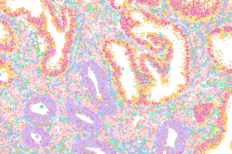
This image reveals the single-cell spatial transcriptomic map of the human endometrium (the inner lining of the uterus), collecting during the “receptive phase” of the menstrual cycle—the brief window when the endometrium is primed for embryo implantation.
This colorful image maps the human endometrium at the single-cell level, with each color representing a different cell type or gene within the uterus’s lining. Yellow and dark pink represent the epithelial glandular cells— specialized epithelial cells that form glands to secrete various substances to prepare for and facilitate the embryo implantation and maintain pregnancy. Purple shows the basal epithelial cells, which contain progenitor cells located at the base of the endometrium. They are anchored to the basement membrane and serve as a source of new cells for the epithelium. The light pink, green and light and dark blues on the image show fibroblasts, which are cells that form and maintain connective tissue.
The dots on the image show different genes: orange (MUC1), pink (EPCAM), red (MME), and yellow (THY1).
Researchers in the Courtois Lab at The Jackson Laboratory, in collaboration with the Department of Obstetrics and Gynecology at UConn Health, study how this crucial stage can be disrupted by endometriosis—a reproductive and systemic disease that affects at least 1 in 10 women. Endometriosis is frequently linked to infertility, yet the biological mechanisms behind this connection remain poorly understood. By examining the endometrium’s cellular and molecular landscape, the team hopes to uncover how endometriosis alters tissue receptivity and impacts fertility.
How was this image captured?
This image was generated using the Xenium platform (10x Genomics), available at the JAX Single Cell Biology Lab (SCBL). The assay detects up to 5,100 genes directly in intact tissue through probe-based cyclic imaging. When paired with antibody-based protein detection—either on Xenium or the complementary Phenocycler Fusion 2.0 (Akoya) platform at SCBL—it enables true multi-omic phenotyping of tissue architecture.
About the Single Cell Biology Lab
The Single Cell Biology Laboratory (SCBL) at JAX helps scientists explore tissues one cell at a time. Using cutting-edge tools like single cell spatial proteomics, transcriptomics and single-cell sequencing and functional 'omics, the lab reveals how cells interact in both healthy and diseased tissues. Researchers work closely with the lab team, which is made up of talented and highly skilled wet lab and computational members in every part of the process from planning experiments to analyzing data in order to get the most from these powerful technologies. The SCBL, led by Director Elise Courtois and Associate Directors Bill Flynn and Kuo-Chan Hung, contributes to national efforts like Cellular Senescence Network (SenNet) and the Human BioMolecular Atlas Program (HuBMAP), helping map human biology in unprecedented detail.
Learn more

Elise Courtois, Ph.D.
Investigates tools for single Cell and Spatial -omics research. Studies endometriosis etiology, pathobiology, and heterogeneity.
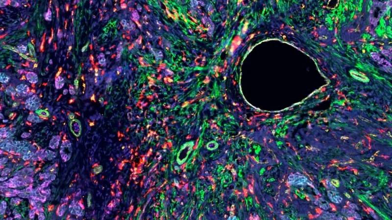
A cellular view of an ovarian tumor
Expert research services at JAX offer unparalleled expertise in mouse models and genetic research, ensuring precision and reliability.
View more