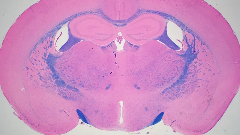
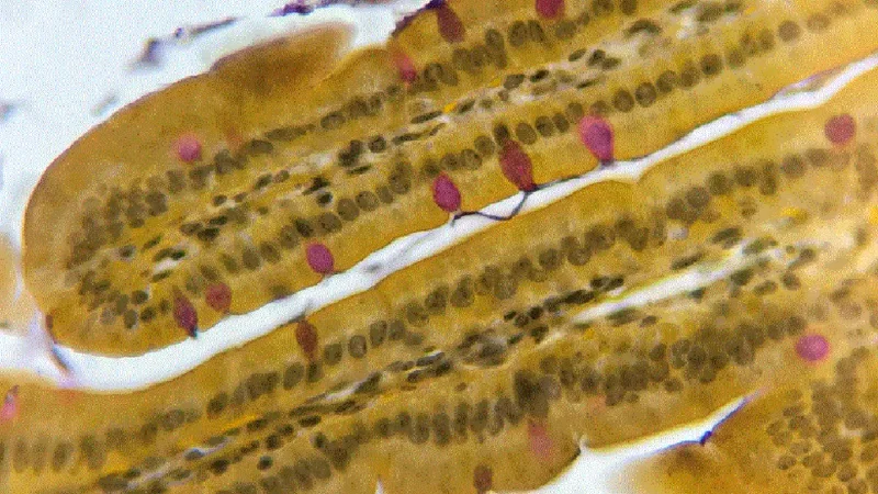
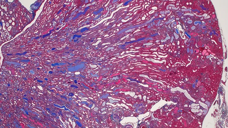
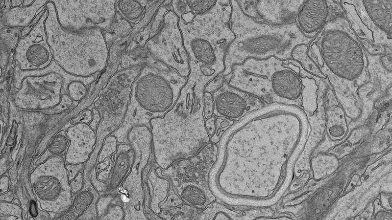
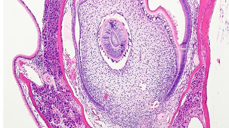
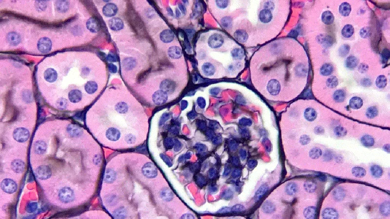
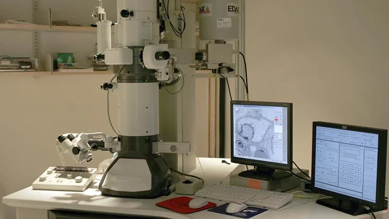
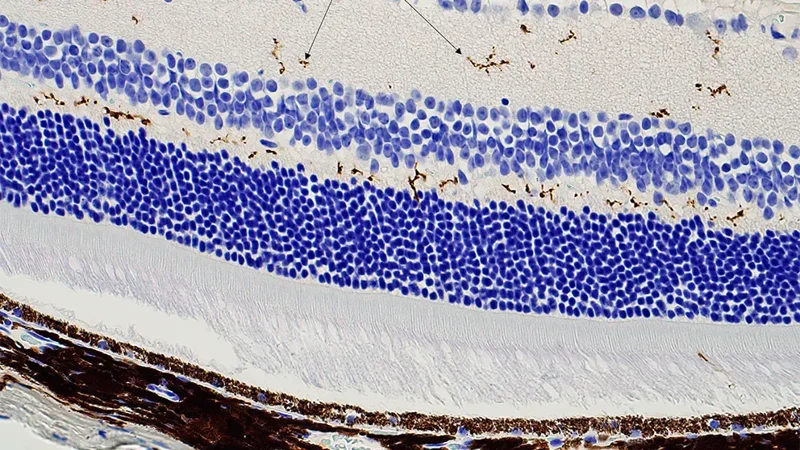
Histopathology Sciences (HPS) provides cutting-edge services enabling analysis of a wide range of anatomical, biochemical, protein, cell, and cell surface characteristics in tissue, blood, urine, and cerebrospinal fluid from various mouse models. The results are critical for numerous and varied phenotyping studies, monitoring disease progression, and determining response to drug treatments or effects of environmental factors. For example, investigators using mouse models of retinopathy rely on the work of HPS to identify lesions in the eye using both Histology and Electron Microscopy Services. The Histology and Clinical Chemistry Services partner with the JAX Diagnostic Program team and Clinical Pathologists to safeguard the health of JAX mouse colonies through the JAX colony monitoring program. The Histology and Electron Microscopy Services can also process fixed human tissue or cultured cell samples, patient-derived xenografts, and non-mammalian vertebrate samples. Our managers and experienced staff are available for consultation. Working together, they provide experimental design support and establish workflows to ensure that investigators can obtain the clearest results for their research.
HPS provides a customized approach to projects and operates on a first-come, first-served basis, ensuring equal access to all services. For projects that require expedited turnaround times, HPS coordinates with investigators to help meet key deadlines. The team focuses on implementing cost-effective approaches so cost is not a barrier to research and scientific progress. Free training is available for multiple methods, allowing users to learn valuable skills while potentially reducing research costs.
Histopathology Sciences consists of the following four Services: