Mapping cellular neighborhoods in a mouse brain
Article | May 8, 2026
For a larger version of this image, click here.
What am I looking at?
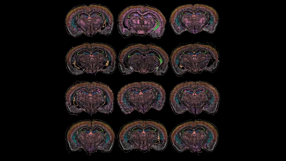
These are coronal slices—or cross-sections—of mouse brains. Each image is a detailed molecular map created with spatial transcriptomics, a technology that reveals where genes are active within intact tissue. Mapping the brain like this helps scientists understand how different cellular neighborhoods interact in health, and how those neighborhoods might degrade or change in diseases like Alzheimer's.
Every color represents a distinct cellular neighborhood defined by unique genetic activity. Neurodegenerative diseases like Alzheimer's don't affect the whole brain equally or at the same time, so the researchers in the O’Connell Lab are trying to pinpoint exactly which cellular neighborhoods are the epicenter of the disease.
The black regions indicate areas where no tissue or detectable gene expression was present. One striking feature is a bright purple cluster that appears minimally in healthy young and aged brains but expands dramatically in diseased tissue. This cluster highlights areas of disease-associated inflammation spreading through the cortex and hippocampus—regions critical for learning and memory.
By comparing the maps across age and genotype, they can track exactly when and where specific cellular functions (like energy metabolism or neuronal signaling) start to break down, allowing us to find new targets for early intervention. The long-term goal is to build a detailed atlas of the aging and diseased brain that could help uncover new targets for earlier diagnosis and intervention.
How was this image captured?
Researchers in the O’Connell Lab analyzed the expression of 247 genes across each brain slice and used an unsupervised machine learning approach called K-means clustering to group regions with similar molecular signatures. Remarkably, the algorithm was not told anything about brain anatomy beforehand. Yet by organizing areas with similar gene activity, it naturally recreated recognizable brain structures, including layers of the cortex, the hippocampus, and white matter tracts.
The tissue was scanned using the 10x Genomics Xenium In Situ platform—an advanced spatial transcriptomics technology that maps where hundreds of genes are turned on at subcellular resolution within intact tissue. After imaging, Surjeet Singh, a postdoctoral associate in the O’Connell Lab developed a custom MATLAB pipeline using unsupervised machine learning to analyze the massive spatial dataset and generate the final color-coded maps.
About the O’Connell Lab
The Kristen O’Connell Lab at JAX studies how aging, metabolism, and neurodegenerative disease affect the brain at the cellular and molecular level. Using advanced mouse models, electrophysiology, single-cell ‘omics, spatial transcriptomics, and spatial mass spectrometry, the lab investigates how individual neurons function, communicate, and change over time in regions involved in memory, cognition, and appetite. The lab has pioneered approaches that link the electrical activity of single neurons with their unique molecular profiles, helping researchers better understand how neuronal identity and function are connected. In parallel, the team is building a spatial multiomic atlas of the brain to map how proteins, lipids, and cellular neighborhoods shift during aging and diseases such as Alzheimer’s, with the goal of uncovering new therapeutic targets for cognitive decline and metabolic disorders.
Learn more
The O'Connell Lab at JAX
Understanding the neural control of appetite and how diet and body weight affect the excitability of the neurons in key CNS circuits responsible for food intake
View more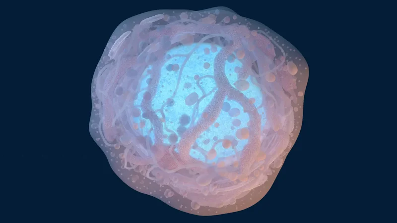
Understanding the structure of a stem cell
This 3D rendering reveals the intricate interior of a human stem cell, centered around a prominent blue nucleus.
View more