The Munnamalai Lab
We investigate how signaling programs instruct in vivo stem cells of the cochlea to differentiate into a highly specialized, organized sensory epithelium.
Principal Investigator
Connect
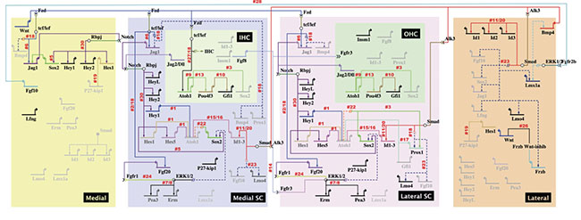
Patterning and gene regulatory network underlying Wnt and Bmp signaling
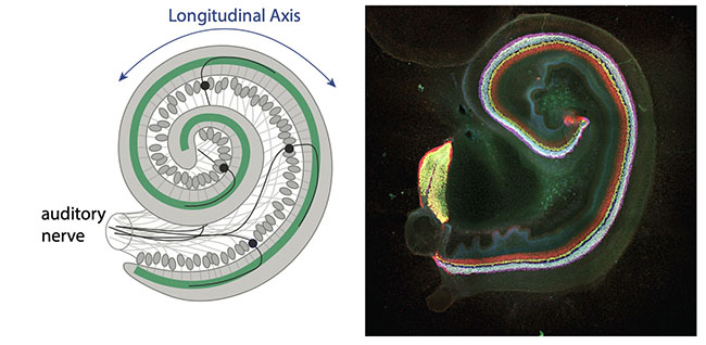
The goal of the lab is to investigate how cell decisions are made during development. To investigate this, the model system we use is the mouse auditory organ due to the complex organization of cells. The auditory organ, the cochlea, has a highly specialized organization for auditory function. There are two types of mechanosensory cells that are required for hearing: Inner hair cells detect sound and the outer hair cells amplify sound. The longitudinal axis specifies frequency selectivity. We use in vitro, in vivo (in utero electroporation) molecular and mouse genetic approaches to validate gene networks.
Patterning and dichotomy of the sensory epithelium
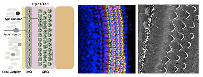
The radial axis determines how the inner and outer hair cells are innervated in the sensory epithelium. The sensory epithelium is known as the organ of Corti. In rodents and humans, there is always one row of inner hair cells (IHCs) to three rows of outer hair cells (OHCs). This 1:3 ratio is essential for proper auditory function.
Asymmetry across the radial axis
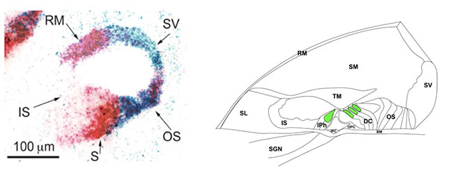
During development, the cochlear duct beings with a very simple structure across the radial axis. These simple epithelia: the Reissner’s membrane (RM), the Stria valscularis (SV), inner sulcus (IS), sensory (S), outer sulcus (OS) will mature to establish a very highly specialized structure and organization. The hair cells are supported by surrounding support cells. We investigate the molecular mechanisms that help specify these cell fate decisions in the cochlea during development.
Counter-gradients to pattern asymmetry
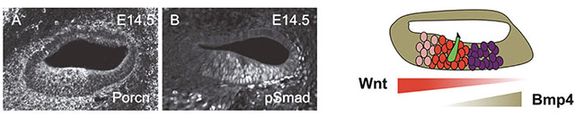
We are interested in two pathways that are important for radial asymmetry: the Wnt and Bmp pathways. We hypothesize that they act in an opposing manner to form counter gradients and hence, asymmetry of the mature cochlea. We are also interested in studying how the asymmetric cellular architecture is established.
Gene Regulatory Networks
Using genomic, perturbation and spatial localization studies, we look to assemble gene regulatory networks to help us model gene interactions underlying cell signaling.
A high-resolution version of this image is available for download.