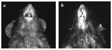
Malocclusion is a common disorder of many strains of laboratory mice and is readily diagnosed by a simple oral examination. Malocclusion occurs in mice when the incisors overgrow because the mandibular and maxillary teeth are not normally aligned and do not properly occlude (see Figure 1).
Rodents are especially prone to malocclusion, since their open-rooted incisors continue to grow throughout life. The tooth enamel of rodents is very hard and often orange-yellow due to the incorporation of iron-containing pigments. In the wild, rodent teeth wear naturally through the consumption of hard foodstuffs and gnawing behavior. In the laboratory, these conditions are mimicked by providing the animals with rodent chow formulated to be hard enough to wear the teeth.
The mouse incisor is composed mainly of dentin, with enamel formed only on the labial surface of the tooth. As the tooth wears, the pulp cells produce more dentin so that the pulp of the tooth, where the nerves reside, is never exposed. Eruption of the incisors in young mice occurs between days 10-12 of age. In mice, the normal rate of eruption (which equals the rate of wear, so that the incisors remain a constant size in adult mice) is approximately 2mm/week for the upper incisors and 2.8mm/week for the lower incisors. This results in a turnover of the entire tooth in 35-45 days(1). If teeth are damaged, the rate of growth may increase.
In mice, malocclusion of both the incisors and the rooted molars has been linked to trauma to developing teeth (cage lids, improper handling, fighting, too-hard food). A genetic basis for some cases of malocclusion is suggested by an increased incidence in certain strains (2, 3, 4). Further, malocclusion is known to be under genetic control in laboratory rabbits.
The diagnosis of malocclusion is often delayed until the mouse is permanently impaired from the malnourishment that attends the condition. Undiagnosed or untreated malocclusion may also result in oral and facial abscesses and osteomyelitis as the growing teeth penetrate facial structures. Malocclusion should be the first disorder suspected when a mouse is smaller and thinner than its littermates at weaning. The standard recommendation for mice with malocclusion found at weaning is euthanasia. When the malocclusion is later in onset and due to trauma, oral tumor formation, or other causes, it is possible to manage the treatment of a valuable mouse through a program of regular tooth trimming using blunt-tipped scissors, to prevent inadvertent oral trauma, and adding a provision of powdered diet. Since the pulp is not exposed in mice, tooth trimming is not painful, although the intensive management and handling necessary to implement a tooth-trimming program may be stressful to the animals. When designing a tooth-trimming regimen, the rapid rate of growth of the incisors must be considered.
| Strain | Stock # |
Percent malocclusion |
|---|---|---|
|
C57BL/6J |
000664 |
0.0460% |
|
B6.V-Lepob |
000632 |
0.0401% |
|
C57BLKS/J |
000662 |
0.0890% |
|
BKS.Cg-m +/+ Leprdb |
000642 |
0.1006% |
|
BALB/cJ |
000651 |
0.0018% |
|
NOD/LtJ |
001976 |
0.0083% |
|
NOD.CB17-Prkdcscid/J |
001303 |
0.0044% |
|
BALB/cByJ |
001026 |
0.0015% |
|
C3H/HeJ |
000659 |
0.0047% |
|
SJL/J |
000686 |
0.0019% |
|
DBA/2J |
000671 |
0.0130% |
References
- Zegarelli EV. Adamantoblastomas in the Slye stock of mice. American Journal of Pathology 1944; 20:23-87.
- Miller WA. Genetic traumatic occlusion in the mouse. J Periodontal Res 1977; 12:64-72.
- Peters H and Balling R. Teeth. Where and how to make them. Trends in Genetics 1999; 15:59-65.
- Petznek H, Kappler R, et al. Reduced body growth and excessive incisor length in insertional mutants mapping to mouse Chromosome 13. Mammalian Genome 2002; 13:504-509.